MAKE A MEME
View Large Image

| View Original: | Pacemaker_NIH.jpg (480x404) | |||
| Download: | Original | Medium | Small | Thumb |
| Courtesy of: | commons.wikimedia.org | More Like This | ||
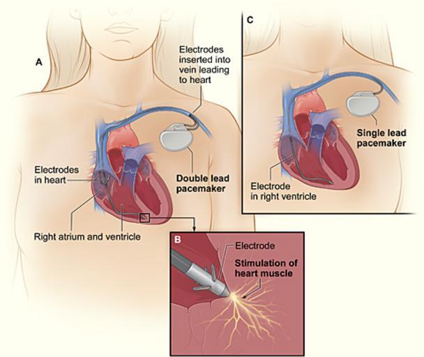
| Keywords: Pacemaker NIH.jpg en The image shows a cross-section of a chest with a pacemaker Figure A shows the location and general size of a double-lead or dual-chamber pacemaker in the upper chest The wires with electrodes are inserted into the heart's right atrium and ventricle through a vein in the upper chest Figure B shows an electrode electrically stimulating the heart muscle Figure C shows the location and general size of a single-lead or single-chamber pacemaker in the upper chest 2013-11-12 22 58 18 National Heart Lung and Blood Institute NIH National Heart Lung and Blood Institute NIH PD-USGov Uploaded with UploadWizard Pacemakers Medical diagrams | ||||