MAKE A MEME
View Large Image

| View Original: | Nicoletti assay 2.png (389x228) | |||
| Download: | Original | Medium | Small | Thumb |
| Courtesy of: | commons.wikimedia.org | More Like This | ||
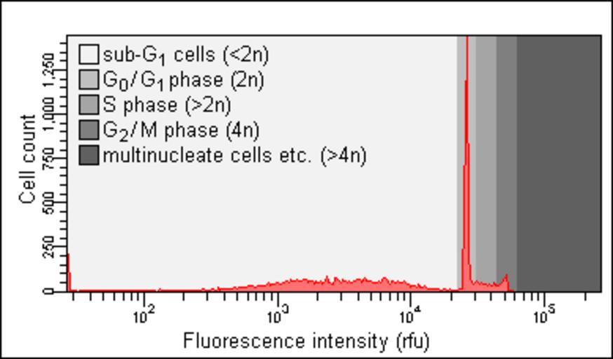
| Keywords: Nicoletti assay 2.png w Histogram of flow cytometry analysis of apoptotic Jurkat cells stained with a fluorescent dye that stains DNA quantitatively that is in proportion to its amount in the cell Five different classes of cells can be identified by the presence of absence of cell count peaks Cells with a DNA content of <2n sub- G0 phase G<sub>0</sub> / G1 phase G<sub>1</sub> These cells are usually the result of apoptotic DNA fragmentation Cells with a DNA content of 2n cells in the G<sub>0</sub>/G1 phase Cells with a DNA content of >2n but <4n cells in the S phase Cells with a DNA content of 4n cells in the G2 phase G<sub>2</sub> Cells with a DNA content of >4n Such cells are usually multinucleate that is they contain multiple cell nucleus nuclei or otherwise aberrant Determining the amount of sub-G<sub>0</sub>/G<sub>1</sub> cells is a method of identifying and to some extent quantifying apoptosis “ note the sub-G<sub>0</sub>/G<sub>1</sub> which is increased as compared to a histogram of healthy cells see images below This method is often referred to as the Nicoletti assay in honour of its inventor the Italian physician Ildo Nicoletti 2012-07-11 own Shinryuu Shinryuu File Nicoletti assay 1 png File Nicoletti assay 3 png Flow cytometry Apoptosis | ||||