MAKE A MEME
View Large Image

| View Original: | Cathepsin K 1TU6.png (1000x719) | |||
| Download: | Original | Medium | Small | Thumb |
| Courtesy of: | commons.wikimedia.org | More Like This | ||
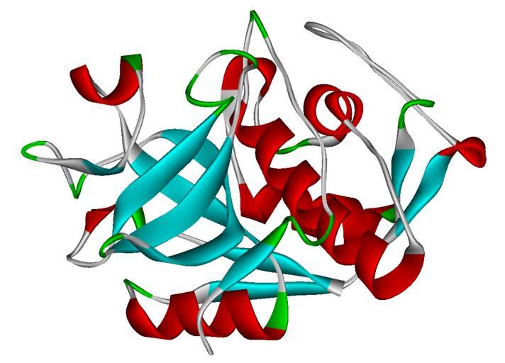
| Keywords: Cathepsin K 1TU6.png Ribbon diagram of human cathepsin K colored by secondary structure <br>Created using http //www accelrys com/products/downloads/ds_visualizer/index html Accelrys DS Visualizer Pro 1 6 and GIMP From PDB entry http //www rcsb org/pdb/cgi/explore cgi pdbId 1TU6 1TU6 2007-03-11 Own work Hydrolases | ||||