MAKE A MEME
View Large Image

| View Original: | Blob sign of ectopic pregnancy.svg (800x345) | |||
| Download: | Original | Medium | Small | Thumb |
| Courtesy of: | commons.wikimedia.org | More Like This | ||
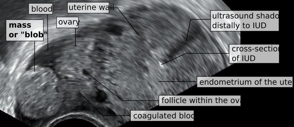
| Keywords: Blob sign of ectopic pregnancy.svg The otherwise healthy 31 year old woman had begun to feel symptoms of pregnancy including breast tenderness A urine pregnancy was positive However three months prior she had a Mirena intrauterine device inserted which thus made the probability of an intrauterine pregnancy very unlikely The vaginal ultrasonography showed the image as displayed including a spherical mass or blob close to the right ovary The intrauterine device was in optimal location within the uterine cavity There was no trace of a gestational sac within the uterus Serum hCG was 1254 IU/L at which level any intrauterine pregnancy is usually seen with the ultrasonography device A cross-section of the Mirena is shown within the uterine cavity leaving an ultrasound shadow distally to it thumb Macroscopic histopathology the fallopian tube and ectopic pregnancy with no distinguishable fetal parts A diagnosis of ectopic pregnancy was made Laparoscopy was performed and showed a fallopian tube that was swollen and bluish as by circulatory obstruction There was 40-50 milliliters of fluid within the rectouterine pouch and around the uterus The affected tube was removed by salpingectomy The postoperative phase was uncomplicated 2014-05-09 own Mikael Häggström thumb left png-format cc-zero Uploaded with UploadWizard Vaginal ultrasonography Ectopic pregnancy | ||||